Last Updated on August 19, 2026
“Time is muscle”
Introduction
Clinical spectrum of ACS

Anatomy-related
• Left anterior descending (
LAD
Complete LAD occlusion (anterior STEMI) = ’widow maker’ 20% of cases of inferior wall of LV infarct are due to left circumflex artery occlusion, i.e. left dominance; 80% of cases of inferior wall of LV infarct are due to RCA occlusion
• Circumflex branch of LCA: posterior wall of LV;
inferior wall of LV
• Right coronary artery: posterior and inferior wall of LV; posterior part of septum; right ventricle; SA node; AV node
Differential diagnoses
• Aortic dissection
Presentation / History Taking
Features of chest pain suggesting ACS
• S: retrosternal Always ask whether the chest pain is at rest / during exertion. Chest pain at rest highly suggestive of ACS instead of stable angina
• C: pressure
• R: jaw / left shoulder / arms
• A: relieved by nitrates within minutes
• T: 5 – 15 mins
• E:
at rest
• S: nausea / vomiting / palpitations / diaphoresis
Gender difference in presentation
• Women likely to present with pain between the shoulder blades / nausea or vomiting / shortness of breath
• Men likely to present with chest pain / diaphoresis
Further reading: https://www.ahajournals.org/doi/10.1161/JAHA.119.014733
Risk factors
• Age >65 Parent(s) / sibling(s)
• Smoking (current / stopped <3 months)
• Diabetes mellitus
• Hypertension
• Hypercholesterolaemia
• Obesity (BMI >30)
•
Family Hx
Others:
• Medications e.g. letrozole
Physical Examination
- Vital signs
-
JVP
×
• Raised JVP ≈Killip 2 MI
- Cardiac
-
Murmur
×Murmur
-
S3 gallop
×
• S3 gallop ≈Killip 2 MI
-
Murmur
- Chest
- To check for any
lung crepitations
×
Lung crepitations ≈pulmonary oedema (Killip 2 and above)
- To check for any
lung crepitations
Scoring
•
HEART score
• Use to predict 6-week risk of major adverse cardiac event TIMI Score for Unstable Angina / NSTEMI Used to predict risk at 14 days of: all-cause mortality, new or recurrent MI, or severe recurrent ischemia requiring urgent revascularization Mnemonics: AMERICA Killip Classification in Acute Myocardial Infarction • Killip Class 1: No signs of left ventricular failure • Killip Class 2: Elevated JVP, Bibasal crepitations, S3 gallop • Killip Class 3: Acute pulmonary oedema • Killip Class 4: Cardiogenic shock
• ≥21 years old, with symptoms suggestive of ACS
•
TIMI
• Age
• Marker (cardiac marker)
• ECG
• Risk factors
• Ischaemic pain (≥2 episodes in 24 hrs)
• Coronary artery stenosis
• Aspirin used in last 7 days
•
Killip
Investigations
Bedside
• ECG
• Capillary blood glucose
Laboratory
• Full blood count
• Renal profile
• LFT, AST
•
Cardiac enzymes
• Coagulation profile
Imaging
• Chest X-ray
ECG in ACS
Cut-off point
ST Elevation
V2 – V3
• Male <40 years old: >1/4 small box
• Male ≥40 years old: ≥2 small boxes
• Female: ≥1.5 small boxes
V4R
• Male <30 years old: ≥1 small box
• Others: ≥half small box
Posterior leads (V7 – V9)
• Male <40 years old: ≥1 small box
• Others: ≥half small box
Other leads
• ≥1 small box
ST Depression
More than half small box
Concordant changes / Contiguous leads
• Inferior MI: II, III, aVF If only leads I and aVL ST elevation, then the MI is high lateral
•
Lateral
Reciprocal changes
Example: Leads I, aVL Leads II, III, aVF
• ST elevation at leads II, III, aVF with ST depression at leads I, aVL strongly suggests (inferior) STEMI
• In
lateral STEMI
=====
STEMI equivalent
• Hyperacute T wave • ST depression V1 – V3 • ST elevation in aVL & V2 • May suggest left main stem occlusion / triple vessel disease de Winter syndrome • Rare but severe [caption id="attachment_1088" align="alignnone" width="389"] Further reading Wellens Syndrome 2 patterns [caption id="attachment_1084" align="alignnone" width="402"] Notes Significance Further reading
•
Posterior MI
• Prominent R wave in V1 – V2
• Upright T in V1 – V3
•
D1 LAD occlusion
• Upright T in aVL and V2
• ST depression & inverted T in leads III & aVF
•
aVR ST elevation with reciprocal changes in concomitant more than 6 leads
•
de Winter syndrome
• Upsloping ST depression (more than 1 mm) with peaked T wave in precordial leads, typically V1 - V4; Reciprocal ST elevation at aVR
• Suggests left anterior descending artery (LAD) completely occluded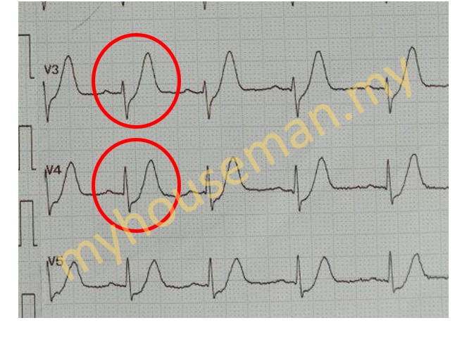 de Winter syndrome[/caption]
LTFL
•
Wellens syndrome
Definition
• Biphasic or deeply inverted T waves in V2-3 PLUS
• History of recent chest pain (now resolved)
Pattern A - Biphasic T waves
Pattern B - Deeply & symmetrical inverted T waves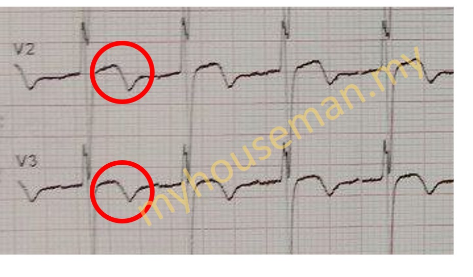 Wellens syndrome - Pattern A[/caption]
• Patient may have normal /slightly elevated serum cardiac markers
• The amplitude of inverted T wave is increased[2]
• Highly specific for critical stenosis of the left anterior descending artery (LAD)
• Extremely high risk for extensive anterior wall MI within subsequent days - weeks
1. LTFL
2.https://www.sciencedirect.com/science/article/pii/S0022073621001187
Management of ACS
Mnemonic: MONA
- Morphine for pain relieve
- IV Morphine 2 – 5 mg over 5 minutes, then PRN (15 minutes intervals)
- +IV Maxolon 10 mg stat and PRN/TDS
- Oxygen supplementation if SPO2 <95% or PaO2 <60 mmHg
- Nitrates
- Sublingual GTN 0.5 mg (maximum 3 times, 5 minutes intervals)
- Antiplatelets
- T Aspirin 300 mg stat (if not given earlier)
- T Clopidogrel 300 mg stat (if not given earlier; if age >75 years old, 75 mg stat)
- Alternatively, if available, T Ticagrelor 180 mg stat can be given if going for primary PCI
Management of STEMI
Assessment of bleeding risk
Contraindications to thrombolysis | ||||
Risk of intracranial haemorrhage | • History of
intracranial bleed
× Intracranial bleed Intracranial bleed (ICB) • History of ischaemic stroke within 3 months • Known structural cerebral vascular lesion (e.g. ateriovenous malformation) • Known intracranial neoplasm | |||
Risk of bleeding | • Active bleeding / bleeding diathesis (excluding menses) • Suspected aortic dissection • Significant head trauma within 3 months | |||
Risk of intracranial haemorrhage | • History of ischaemic stroke >3 months • Severe uncontrolled HTN on presentation (BP >180 / 110 mmHg) • Chronic, severe uncontrolled hypertension | |||
Risk of bleeding | • Current use of anticoagulant in therapeutic doses (INR >2) or
DOAC
Abbreviation: DOAC Direct oral anticoagulant Examples: Apixaban, Rivaroxaban • Recent major surgery <3 weeks • Recent internal bleeding (GI / urinary tract haemorrhage) • Active peptic ulcer • Non-compressible vascular puncture • Traumatic / prolonged CPR >10 minutes | |||
Others | • Pregnancy • Prior exposure (>5 days and within 12 months of first usage) to
streptokinase
Streptokinase Streptokinase | |||
• Large infarcts • Anterior infarcts • Hypotension / cardiogenic shock • Significant arrhythmias • Elderly • Post-revascularization (post-CABG / post-PCI) • Post-infarct angina | ||||
Reperfusion strategy
×Establishing time intervals to reduce total ischaemic time AND achieve timely early reperfusion
Establishing time intervals to reduce total ischaemic time AND achieve timely early reperfusion
Thrombolysis:
Streptokinase
Streptokinase
COROS / PCI / Cardiac angiogram
Successful reperfusion
- Resolution of chest pain
- Early return of ST elevation to isoelectric line // decrease in height of ST elevation by 50% (in the lead that records highest ST elevation) within 60 – 90 minutes of initiation of fibrinolytic therapy
- Restoration and/or maintenance of haemodynamic and/or electrical stability
- Early peaking of CK & CK-MB level
Medications in STEMI
Antiplatelets
Double antiplatelet (DAPT)
- T Aspirin 300 mg loading dose (given on arrival), then 75 –
100 mg OD
×
e.g. T Cardiprin 100 mg OD
- P2Y12 inhibitor: T Clopidogrel 300 mg or
75 mg
loading dose (given on arrival), then 75 mg OD×
For patients aged more than 75 years old
Duration of DAPT
Balance between bleeding risk vs ischaemic/thrombotic risk
- Post fibrinolysis: 1 month to 1 year
- Post PCI: 6 months to 1 year
Anti-thrombotic therapy (Anti-coagulant)
Injectable anticoagulant
For patients who received fibrinolytic therapy without undergoing PCI // Not receiving fibrinolytic therapy & PCI
(i) Enoxaparin (Clexane)
– <75 years: IV Clexane 30 mg stat, then SC Clexane 1 mg/kg BD for 8 days or until hospital discharge
– ≥75 years: SC Clexane 0.75 mg/kg BD for 8 days or until hospital discharge
(ii) Fondaparinux (Arixta)
– SC Fondaparinux 2.5 mg OD for 8 days or until hospital discharge
(iii) Unfractionated heparin (UFH)
– IV Heparin 60 units/kg bolus (max 4000 units), then IVI Heparin 12 units/kg/hr (max 1000 units/hr), target APTT 1.5 – 2.5 x control, for 48 hours
Management of NSTEMI
Management of UNSTABLE ANGINA
Treatment Modalities in ACS
Percutaneous coronary intervention (PCI)
Thrombolysis
Common medications used u0026amp; doses
Related Posts
• Chest pain [open]
Clinical Questions
--- This content is for premium members only. ---