Last Updated on August 19, 2026
Introduction
• Pneumothorax = presence of air in pleural space
Classification
• Closed pneumothorax
• Open pneumothorax
• Tension pneumothorax
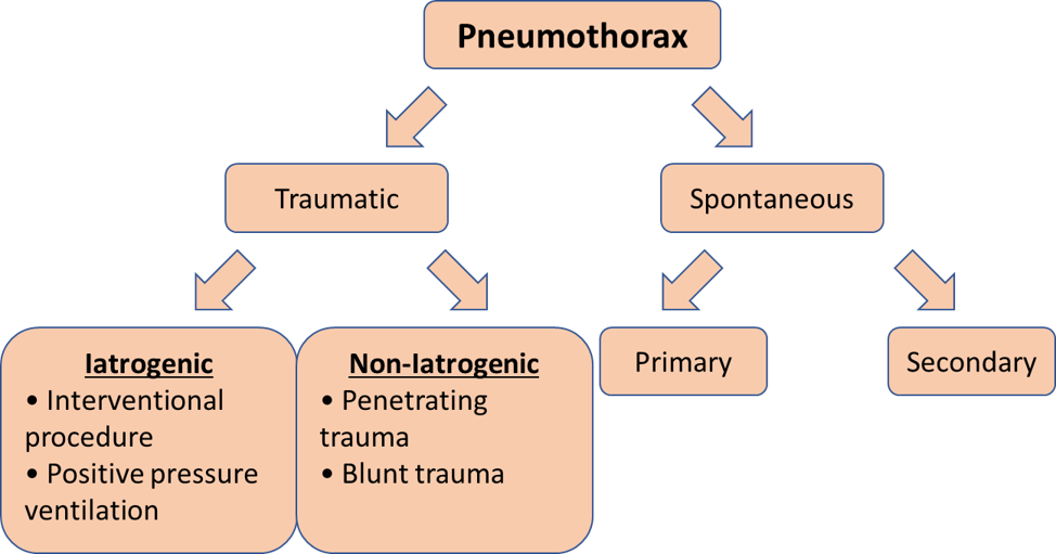
Traumatic pneumothorax
Examples of interventional procedures that cause pneumothorax
• Intercostal nerve block
• Chest aspiration
• Trachiobronchial biopsy
• Needle aspiration
• Lung biopsy
Secondary pneumothorax
Causes:
• COPD
• Asthma
• Tuberculosis
• Pleural malignancy
• Marfan’s syndrome
• Bacterial pneumonia
• Rheumatoid lung disease
Presentation
•
Chest pain
Characteristics:
• On the side of pneumothorax
• Sharp & pleuritic
• May have SOB
Clinical Features
Physical examination
(i) Lungs Especially in tension pneumothorax In tension pneumothorax, air accumulates between chest wall and lung, increasing pressure in the chest → reduced amount of blood returned to the heart → jugular vein distension Remember, tension pneumothorax is an example of obstructive shock
• Reduced chest movement on the affected side
• Hyper-resonance of affected side
• Decreased breath sounds of affected side
(ii)
Other findings
•
Neck vein distension
• Subcutaneous emphysema of the chest wall / neck
• Trachea deviated away from affected side
•
Hypotension
Management
• High flow-mask
Management of open pneumothorax
Temporary measure:
• Application of three-way occlusive dressing to the wound [Video]
Followed by:
• Chest tube insertion
Management of tension pneumothorax
• Chest tube insertion Safety Triangle Why safety triangle is important? Notes:
=====
💡Notes
• If chest tube can be inserted immediately, no need to perform chest needle decompression prior to chest chest tube insertion
• Needle decompression for adult can be performed at
safety triangle
 Borders of safety triangle
• Anterior border: Lateral border of pectoralis major
• Lateral border: Lateral border of latissimus dorsi
• Inferior border: 5th intercostal space
• A site with minimised risk of internal organ perforation during chest tube insertion
• For maximum exposure of safety triangle, externally rotate the ipsilateral arm with hand above the head
Management of spontaneous pneumothorax
• Figure 2: Flowchart of management of spontaneous pneumothorax by British Thoracic Society
Points towards secondary spontaneous pneumothorax
• Age ≥50
• Smoking
• Evidence of underlying lung disease on exam / imaging
Primary pneumothorax with
Chest tube removal
Indications
- Lung is
fully expanded
×
• Lungs auscultation: equal air entry bilaterally
• CXR post chest tube insertion: no pneumothorax - No evidence of
ongoing air leak
×
evidenced by bubbling in the underwater seal
Preparation for chest tube removal
- Chest tube may be clamped for few hours prior to removal
- Observe for evidence of pneumothorax
- If pneumothorax recurs, chest drain can be unclamped, and chest drain cannot be removed first
Further reading
Related Posts
Q&A
Take Home Message
1. (Tension) Pneumothorax is a potentially fatal medical emergency without immediate treatment
2. (Tension) Pneumothorax is a clinical diagnosis. Do not wait until CXRavailable, which would delay treatment×CXRAbbreviation: CXR